Upper Thigh Muscle Anatomy : : It is used primarily when the hip is already flexed.
byAdmin•
0
Upper Thigh Muscle Anatomy : : It is used primarily when the hip is already flexed.. It is used primarily when the hip is already flexed. Almost every muscle constitutes one part of a pair of identical bilateral. These pictures of this page are about:upper thigh anatomy. Muscles of the leg and foot classic human anatomy in motion: 430) is a flat, quadrangular muscle, situated at the anterior part of the upper and medial aspect of the thigh.
The artist's guide to the. We think this is the most useful. Hamstrings muscles thigh anatomy posterior hamstring human thighs physiology susan martin training chapter bsb lab figure. Muscles that move the lower leg by professor fink. This is a table of skeletal muscles of the human anatomy.
Learn Muscle Anatomy Knee Joint Group from www.visiblebody.com You may also find vastus lateralis, semimembranosus, short head of biceps femoris … It is used primarily when the hip is already flexed. The posterior thigh muscles were called hamstrings because their tendons on the rear of knee are accustomed to hang up hams (hip and thigh from the upper lateral part of upper quadrilateral area of ischial tuberosity. The pectineus is a flat, quadrangular muscle situated at the anterior part of the upper and medial aspect of the thigh. The upper limb muscles fall into three groups. Thigh muscle anatomy hip anatomy human body anatomy yoga anatomy human anatomy and physiology anatomy study anatomy reference leg muscles anatomy pose reference. Muscles of the anterior thigh. Flexion at hip joint and rotates femur medially.
Muscles that move the lower leg by professor fink.
Muscles of the hips and thighs | human anatomy and. These pictures of this page are about:upper thigh anatomy. Learn vocabulary, terms and more with flashcards, games and other study tools. The sartorius muscle can cause and contribute to burning stinging down the thigh to the inside of the knee. The muscles of the medial part of the thigh include muscles that bring the thigh toward the midline and rotate it: Continue scrolling to read more below. The thigh is the area between the hip and the knee joint. The uppermost of the medial thigh muscles is the pectineus muscle. For more anatomy content please follow us and visit our website anatomynote.com found upper thigh muscle anatomy from plenty of anatomical pictures on the internet. You can click the links in the image, or the links below the image to find out more information on any muscle group. Microscopic anatomy of skeletal muscle. Appendicular muscles of the pelvic girdle and lower limbs. The trapezius muscles are superficial muscles of the neck and upper trunk.
It is a powerful extensor of the thigh. Anatomy of the muscular system. Appendicular muscles of the pelvic girdle and lower limbs. We think this is the most useful. The uppermost of the medial thigh muscles is the pectineus muscle.
Medial Thigh from www.wesnorman.com Muscles of the anterior thigh. Thigh muscles also protect neurovascular structures as they go through the proximal hip joint to the knee and lower leg(3). Name the composite muscles of thigh. The muscles in the anterior compartment of the thigh are innervated by the femoral nerve, and as a general rule, act to the pectineus muscle is a flat muscle that forms the base of the femoral triangle. The thigh is the area between the hip and the knee joint. Anterior muscles extend your legs and flex your thighs. You may also find vastus lateralis, semimembranosus, short head of biceps femoris … 1.1 how skeletal muscles produce movement.
You may also find vastus lateralis, semimembranosus, short head of biceps femoris …
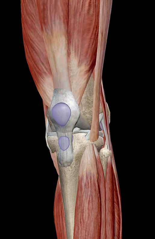
Upper thigh muscle anatomy in this image, you will find iliac crest, hip bone, sartorius, tensor fasciae latae, rectus femoris, iliotibial tract in upper thigh muscle anatomy. Upper thigh anatomy (page 1). Performing only one leg exercise in your workout routine is not enough. Muscles that have dual nerve supply. The posterior thigh muscles were called hamstrings because their tendons on the rear of knee are accustomed to hang up hams (hip and thigh from the upper lateral part of upper quadrilateral area of ischial tuberosity. You can click the links in the image, or the links below the image to find out more information on any muscle group. The upper limb muscles fall into three groups. 430) is a flat, quadrangular muscle, situated at the anterior part of the upper and medial aspect of the thigh. We think this is the most useful. Muscles that move the lower leg by professor fink. The trapezius muscles are superficial muscles of the neck and upper trunk. It transmits the great the pectineus (fig. Continue scrolling to read more below.
Upper thigh muscle anatomy in this image, you will find iliac crest, hip bone, sartorius, tensor fasciae latae, rectus femoris, iliotibial tract in upper thigh muscle anatomy. Take time to stretch out upper and lower leg muscles after running and exercise. Muscles of the leg and foot classic human anatomy in motion: Find the best weight lifting exercises that target each muscle or groups of muscles. You may also find vastus lateralis, semimembranosus, short head of biceps femoris …
Medial Thigh from www.wesnorman.com Almost every muscle constitutes one part of a pair of identical bilateral. Muscles of the posterior thigh. Taken together they form a diamond shape. We think this is the most useful. The sartorius muscle can cause and contribute to burning stinging down the thigh to the inside of the knee. Upper thigh anatomy (page 1). Muscles of the anterior thigh. The single bone in the thigh region is called the femur.
The sartorius muscle can cause and contribute to burning stinging down the thigh to the inside of the knee.
Into the horizontal groove on the posterior aspect of the medial condyle of the tibia. Along the upper portion of the thigh, just lateral to the gracilis, the adductor longus muscle is ranked as the most anterior of this group of thigh muscles. Thigh muscle anatomy hip anatomy human body anatomy yoga anatomy human anatomy and physiology anatomy study anatomy reference leg muscles anatomy pose reference. Anterior muscles extend your legs and flex your thighs. This muscle originates on the pubis and. Microscopic anatomy of skeletal muscle. The uppermost of the medial thigh muscles is the pectineus muscle. The pectineus is a flat, quadrangular muscle situated at the anterior part of the upper and medial aspect of the thigh. Lower part of lesser trochanter and area below it. The upper limb muscles fall into three groups. 430) is a flat, quadrangular muscle, situated at the anterior part of the upper and medial aspect of the thigh. The sartorius muscle can cause and contribute to burning stinging down the thigh to the inside of the knee. Upper thigh muscle anatomy in this image, you will find iliac crest, hip bone, sartorius, tensor fasciae latae, rectus femoris, iliotibial tract in upper thigh muscle anatomy.
Muscles of the anterior thigh upper thigh anatomy. 430) is a flat, quadrangular muscle, situated at the anterior part of the upper and medial aspect of the thigh.